How does Laparoscopic surgery work?
In order to gain entry into the abdomen small incisions are made through the skin. The incisions range from 3mm to 12mm (0.11 to 0.5 inches) depending on what instruments are required to safely perform the procedure. On occasion one incision may have to be elongated to retrieve a specimen (i.e. fatty tissue, hernia sac).
Through the skin incisions a plastic/ metal tubes (referred to as ports or trocars) are introduced into the abdomen through the subcutaneous fat and muscle layers of the abdominal wall. Once entry has been achieved, the camera (laparoscope), instruments, blood vessel sealing devices, surgical staplers and a myriad of other tools can be exchanged with ease.
In order to create a “working space” the abdominal cavity is insufflated or distended with a non-toxic, nonflammablegas (CO2) through one of the ports placed into the abdominal cavity. CO2 (carbon dioxide) is the end product of metabolism in all living organisms. In mammals, the overwhelming majority of CO2 is expelled through breathing and the kidneys. In fact, every time we exhale we are “blowing off” CO2. Almost all CO2 used during laparoscopy is absorbed and eliminated by our body within 48 hours.
Once access and exposure has been achieved the surgical procedure can begin and often follows the exact same steps and principles as the open procedure.
Laparoscope (camera)
 What is commonly referred to as the laparoscope is a combination of three devices, the lens, camera and a fiber optic cable system. The telescopic rod lens system is housed in a long telescope that is then connected to a camera. The camera is connected to an image processor that will then project the image onto a monitor. A digital laparoscope houses the image processor at the end of the telescope and precludes the need for a rod lens system. All systems require a fiber optic cable system hooked up to a “cold” light source (Xenon or Halogen) to illuminate inside the abdominal cavity.
Trocars (Ports)
For the surgeon to access the abdominal cavity, long thin tubes (trocars/ports) are passed through the muscles of the abdominal wall after making a small skin incision. The port also provides a way to fill the abdomen with CO2 in order to distend the abdominal cavity creating a working space. The laparoscope and instruments can then be passed through the ports to begin the procedure. Many different instruments are used throughout any given procedure. The ports allow easy exchange of instruments.
General Instrumentation
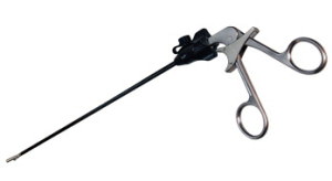
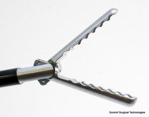
In general, the instruments used in laparoscopic surgery are a combination of three parts: handle, shaft and actuator. There are several handles that are available and surgeon ease and comfort generally determines what handles are used. Regardless of design the handles are opened and closed to control the actuator. The shaft is a long tube that connects the handle to the actuator. The length of the shaft is generally 30-45 cm. Instruments that utilize electrocautery must have an insulated shaft to prevent arcing and burns to tissue in contact with the shaft. The actuator is at the end of the shaft and is generally what makes one instrument different than another. The actuator may be a grasper, scissors, biopsy forceps amongst many others.
Graspers
Graspers allow the surgeon to hold and manipulate organs or tissue. It is used like forceps in open surgery. Graspers are used in most laparoscopic procedures.
Dissector
Dissector has a fine tip and is used for very delicate dissection. The angle of the tip varies from straight to a slight curve to a right angle (90 degrees).
Scissors
Scissors are commonly used to cut tissue and sutures. The scissors can also be used for fine dissection.
Needle driver
Needle drivers are used to sew and tie sutures inside the abdominal cavity, still through small incisions. This is considered to be one of the hardest laparoscopic skills to master, and is one of the more recognizable instruments used by laparoscopic surgeons.
Cautery/Tissue sealing devices
There are several instruments that are used to cauterize and divide tissue available to the surgeon. Some forms utilize high frequency vibration to generate heat in order to keep tissue from bleeding.
Clip applier
The clip applier is used to ligate or occlude a blood vessel or other hollow tube. The clip applier is most commonly used in gallbladder surgery to ligate the cystic artery and cystic duct. This prevents bleeding or leakage of bile after the gallbladder is removed. The clips are made of titanium.
Stapling devices
Stapling devices are used to divide hollow organs or blood vessels. Staplers are infrequently used in hernia surgery. The stapler locks 6 rows of staples into the tissue and a blade is passed through the center of 3rd and 4th row simultaneously sealing and dividing. The length of the staple line as well as staple height varies between different cartridges. The surgeon will decide what staple cartridge to use dependent on the procedure. The staples are made of titanium.


